automatic brightness control in fluoroscopy
This is a function of Fluoroscopy, which controls the overall performance of the fluoroscopic image by automatically adjusting kVp, mA or both. By this, the image brightness is kept fixed on the monitor. It is a feed back circuit that measures the light intensity or video camera signal of the output screen.
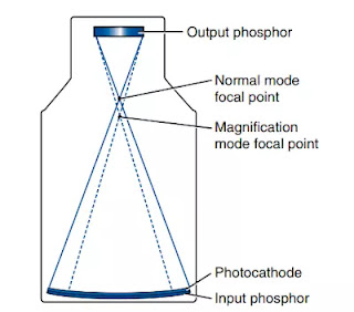
Image A photomultiplier tube or a photocathode is used to monitor the light output of the intensifier tube. When the corresponding brightness changes, it is sent to the generator for adjustment. The generator controls the exposure rate by changing kVp, mA or both. The brightness of the central area of the output screen is measured for this adjustment.
Brightness can be adjusted with both kVp, mA, which affects both contrast and parient dose. The three methods of adjustment are as follows -
- Change of kV at constant mA
- Change of mA at constant kV
- Change of both kV and mA


