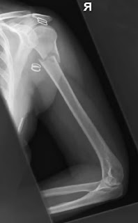
fractured humerus x-ray
1. Humerus Antero-posterior – erect
Position of patient and cassette
Erect Bucky में "35×43"cm कैसेट लगाते है । patient को image receptor के contact में खड़ा करते हैं। तथा patient को affected side की ओर थोड़ा सा rotate करते हैं ताकि shoulder , upper arm तथा elbow का posterior aspect कैसेट के contact में आ जाये । patient position को इस तरह adjust करते हैं की humerus के lateral तथा medial epicondyle cassette से समान दूरी पर हो।Direction and centring of the X-ray beam
2. Humerus lateral erect
Position of patient and cassette
Vertical bucky में "35×43" सेंटीमीटर की कैसेट लगाते हैं AP पोजीशन से पेशेंट को 90 डिग्री पर इस तरह से rotate करते हैं कि arm की लेटरल सरफेस कैसेट के contact में आ जाए। इसके बाद पेशेंट को इस तरह से रोटेट करते हैं कि उसकी arm rib cage से दूर हो जाए लेकिन arm फिर भी कैसेट के कोंटेक्ट में रहे।Direction and centring of the X-ray beam
Collimated horizontal x-ray beam के सेन्टर को shoulder तथा elbow joint के मध्य में इमेज रिसेप्टर के लम्बवत देते हैं।Essential image characteristics
Exposure factor को इस तरह से रखते हैं जिससे कि area ofinterest सही से दिखाई दे।


