Translate

BUY NOTES

BUY NOTES
positioning लेबलों वाले संदेश दिखाए जा रहे हैं. सभी संदेश दिखाएं

radiography positioning notes


radiography positioning


जिस टॉपिक को आप पढना चाहते हो उस पर क्लिक करे 

pdf  की नई अपडेट पाने के लिए whatsapp no. 7891738075


-  Terminology and position of body
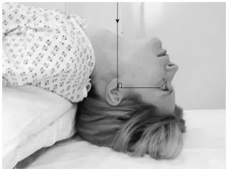
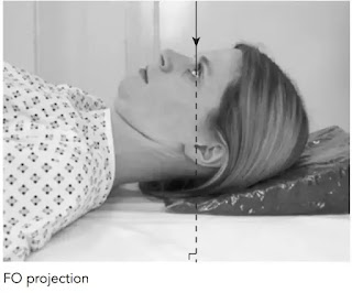
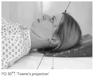
   skull
   chest

   upper limb


   lower limb


   pelvic girdle


  vertebral column 

skull Stenver's or petrous anterior oblique x-ray view in hindi

skull Stenver's or petrous anterior oblique x-ray view in hindi

Position of patient and cassette

यह projection prone पोजीशन में भी किया जा सकता है लेकिन पेशेंट के लिए erect पोजीशन अधिक comfortable रहती है। पेशेंट को vertical bucky की तरफ मुँह करके खड़ा किया जाता हैं। जिस साइड का examination किया जाना है उस साइड के supra-orbital margin के सेन्टर को bucky के बीच मे centred करते हैं। गर्दन को flex करके nose तथा forehead को cassette के contact में रखते है। तथा orbito-meatal line को टेबल लम्बवत रखते हैं। 

जिस अवस्था मे median sagittal plane टेबल से लम्बवत हो उस पोजीशन से पेशेंट के head को examination side की तरफ से इस प्रकार rotate किया जाता  है  जिससे कि median sagittal plane टेबल से 45° के angle पर हो जाए। इससे temporal bone का petrous part कैसेट के समांतर हो जाए। अब थोड़ा गर्दन को इस प्रकार extended करते है जिससे कि orbito-meatal line क्षेतिज से 5 डिग्री angle पर हो जाए। एक 18×24cm साइज की cassette को bucky के अंदर transversely इस प्रकार place करते जिससे कि कैसेट का सेंटर central ray के अनुरुप हो।

skull Stenver's or petrous anterior oblique x-ray view in hindi


Direction and centring of the x-ray beam

Collimated horizontal x-ray beam को 12° cranially angle देते है। तथा इसके सेन्टर को xray cassette से दूर स्थित external auditory meatus तथा occipital protuberance के बीच देते है। temporal के petrous भाग तथा mastoid को examination area में शामिल करते हुए beam को collimate करते है। 

skull Stenver's or petrous anterior oblique x-ray view in hindi


Note -: CT scan  के आने के बाद यह projection बहुत कम किया जाता हैं।

skull Reverse Towne's / OF 30° cranial tilt view in hindi

skull Reverse Towne's / OF 30° cranial tilt view 

Position of patient and cassette

यह projection सामान्यतः erect पोजीशन में लिया जाता है। तथा पेशेंट को bucky तरफ मुँह करके खड़ा किया जाता हैं। हालाँकि यह prone पोजीशन में भी performed किया जा सकता हैं। सबसे पहले patient को nose तथा forehead को bucky table पर रखने को कहा  जाता हैं। पेशेंट के head को इस प्रकार adjust करते हैं जिससे कि median sagittal plane cassette की midline के ऊपर right angle पर रहे । orbito-meatal baseline कैसेट के लम्बवत होनी चाहिए। stability के लिए पेशेंट को bucky के किनारे को पकड़ा सकते है।

skull Reverse Towne's / OF 30° cranial tilt view  in hindi


Direction and centring of the x-ray beam

Central ray को cranially angle देने के लिए इसे orbito-meatal plane से 30° angle पर देते है। collimation field को इस प्रकार सेट करते है जिससे कि सम्पूर्ण occipital bone तथा vertex  parietal bone इस field में शामिल रहे। तथा lambdoidal suture साफ दिखाई देना चाहिए। पेशेंट की आंखों को primary beam से बचाने की कोशिश करते है। skull के lateral skin margins इमेज में शामिल होने चाहिए।

skull Reverse Towne's / OF 30° cranial tilt view  in hindi


Essential image characteristics

skull में किसी भी प्रकार का rotation नही होना चाहिए। sphenoid bone की sella turcica foramen Magnum में  दिखाई देनी चाहिए।

skull Mastoid profile view in hindi

skull Mastoid profile view

Position of patient and cassette

पेशेंट को x-ray table पर supine सुलाते है साथ ही orbito-meatal baseline को table top से लम्बवत रखते है। median sagittal plane टेबल से लम्बवत हो तथा इस पोजीशन से पेशेंट के head को 35° उस तरफ rotate किया जाता है जिस तरफ का examination लेना है। जिससे कि अब median sagittal plane टेबल से 55 डिग्री का angle बनाए। finally पेशेंट के head को transversely इस प्रकार move करते है जिससे कि  mastoid process टेबल की midline में examined हो।

skull Mastoid profile view in hindi


Direction and centering of the x-ray beam

Central ray को caudally angle देने के लिए इसे orbito-meatal plane से 25° के एंगल पर देते है। तथा इसके सेन्टर को जिस तरफ का examination लेना है उस तरफ की mastoid process के बीच देते है। mastoid process के सभी तरफ के area को collimate करते है।

skull Mastoid profile view in hindi


Note -: दोनों तरफ के अंतर को देखने के लिए दोनों तरफ का radiograph लिया जाता हैं। एक छोटे lead marker को collimation area में शामिल करना चाहिए।

skull Mastoid - lateral oblique 25 degree caudad in hindi

skull Mastoid - lateral oblique 25 degree caudad

Position of patient and cassette

पेशेंट को erect bucky की तरफ मुँह करके बैठाया जाता हैं। पेशेंट के head को इस प्रकार rotate करते है जिससे कि median sagittal plane Bucky के समान्तर हो जाए तथा   inter-orbital line bucky के लम्बवत हो जाए। सही पोजीशन प्राप्त करने के लिए शोल्डर को हल्का सा rotate करते हैं। stability के लिए पेशेंट bucky को पकड़ सकता है। cassette के तरफ वाले ear की earpinna को आगे की तरफ fold कर देते हैं जिससे कि यह mastoid region पर superimposed ना हो। एक 18×24cm size की cassette को bucky के अंदर longitudinally place करते हैं। तथा इस cassette का सेन्टर को central ray तथा mastoid process के अनुरूप रखते हैं।

skull Mastoid - lateral oblique 25 degree caudad in hindi


Direction and centring of the x-ray beam

एक 25 degree caudal angulation को काम मे लेते हुए इसके सेन्टर को cassette से दुरस्थित external auditory meatus से 5cm ऊपर तथा 2.5cm पीछे की ओर देते हैं।  examination area को collimate करते है |

skull Mastoid - lateral oblique 25 degree caudad in hindi


Essential image characteristics

यह सुनिश्चित करते हैं कि सभी mastoid air cells इमेज में शामिल हैं । दोनों तरफ की mastoid air cells के अंतर को देखने के लिए दोनों तरफ का radiograph लिया जाता हैं।

skull Jugular foramina / submento vertical 20 degree caudad view in hindi

skull Jugular foramina / submento vertical 20 degree caudad view

Jugular foramina  स्कल के पिछले क्रेनियल fossa में temporal के petrous तथा occipital bone के बीच foramen magnum के दोनों ओर स्थित होता है। इन दोनों foramina की इमेज को svm पोजीशन में 20° caudal एंगल देकर प्राप्त कर सकते हैं।

Position of patient and cassette 

यह प्रोजेक्शन erect या supine पोजीशन में किया जा सकता हैं। यदि पेशेंट अस्थिर रहे तो supine technique उचित रहती हैं। पेशेंट को टेबल की सेन्टर लाइन पर supine position में लेटाते है। पेशेंट के पीठ तथा shoulder को उठाकर उनके नीचे pillow लगा देते है। अब पेशेंट की chin तथा neck को पीछे की दिशा में मरीज की सुविधा अनुसार तब तक झुकाते जाते है जब तक कि skull का vertex x-ray table की सेन्टर लाइन से touch ना हो जाए। अर्थात base line टेबल के समांतर आ जाये एवं median sagittal plane टेबल के लम्बवत आ जाये।

skull Jugular foramina / submento vertical 20 degree caudad view in hindi


Direction and centring of the x-ray beam

एक अच्छे collimated beam का प्रयोग करते हुए central ray को base line के बीच देते हैं । और x-ray beam को लम्बवत से 20° caudal एंगल देते हैं । इस पोजीशन में x-ray beam orbito-meatal plane से 70° डिग्री के एंगल पर होता है।


skull Jugular foramina / submento vertical 20 degree caudad view in hindi


Skull optic foramina view in hindi

Skull optic foramina view in hindi

बोनी ऑर्बिट के पीछे optic foramen में optic canal खुलती है। यह canal median sagittal plane से 35° लेटरल तथा OM line से 35° नीचे open होती हैं। इसलिए इसकी pathology के लिए x-ray beam को इसके बीच से गुजारते है। इसमे comparison के लिए दोनों orbit canal का लिया जाता है। 

Position of patient and cassette

Patient को x-ray table की तरफ मुँह करके खड़ा किया जाता हैं। अब जिस साइड का examination करना है उस साइड check , chin तथा nose को cassette के contact में रखते हैं। orbit के सेंटर ( जिस साइड की orbit का examination किया जाना है ) को cassette या bucky के सेन्टर पर रखते हैं । यह पोजीशन इस प्रकार होनी चाहिए जिससे कि msp टेबल से 35° angle  तथा orbito-meatal baseline horizontal से 35° का एंगल बनाये अर्थात।

Skull optic foramina view in hindi


Direction and centring of the x-ray beam

Collimated horizontal x-ray beam का सेंटर bucky के बीच होना चाहिए। यह बिंदु external auditory meatus से 7.5cm ऊपर तथा 7.5cm पीछे होता है।  एक छोटे lead के side marker को orbital margin के ऊपर place कर सकते हैं।

Skull optic foramina view in hindi


Skull Submento vertical / base of skull / hanging head view in hindi

Skull Submento vertical / base of skull / hanging head view

यह projection skull के base , occipital bone , Atlas के anterior arches  तथा mastoid air cell आदि की pathology देखने के लिए किया जाता हैं। यह प्रोजेक्शन erect या supine पोजीशन में किया जा सकता हैं। यदि पेशेंट अस्थिर रहे तो supine technique उचित रहती हैं।

Position of patient and cassette 

पेशेंट को टेबल की सेन्टर लाइन पर supine position में लेटाते है। पेशेंट के पीठ तथा shoulder को उठाकर उनके नीचे pillow लगा देते है। अब पेशेंट की chin तथा neck को पीछे की दिशा में मरीज की सुविधा अनुसार तब तक झुकाते जाते है जब तक कि skull का vertex x-ray table की सेन्टर लाइन से touch ना हो जाए। अर्थात base line टेबल के समांतर आ जाये एवं median sagittal plane टेबल के लम्बवत आ जाये।

Skull Submento vertical / base of skull / hanging head view in hindi


Direction and centring of the x-ray beam

Central ray को orbito-meatal plane के right angle ( 90° डिग्री ) पर दोनों external auditory meatuses के बीच देते है।

Skull Submento vertical / base of skull / hanging head view in hindi


Essential image characteristics

एक सही प्रोजेक्शन में angle of mandible तथा temporal bone का petrous portions साफ दिखाई देते हैं। middle cranial fossa का foramina   midline के एक तरफ समरूपता से दिखाई देना चाहिए।

Skull Fronto-occipital view in hindi

Skull Fronto-occipital view

Skull के Fronto-occipital projection में same anatomy को देख सकते हैं जैसा कि occipito-frontal projection में दिखाई देती हैं। इसमे orbits तथा frontal bone magnified हो जाती हैं ।  यह प्रोजेक्शन तभी लिया जाता है जब पेशेंट को move नही किया जा सकता हो या supine इमेज लेना बहुत जरूरी हो। सामान्यतः यह प्रोजेक्शन नही लेते हैं क्योंकि इस प्रोजेक्शन  के कारण eye radiation dose बढ़ता है तथा object to film distance ( OFD ) ज्यादा होने के कारण anterior skull structures का resolution कम हो जाता हैं।

Position of patient and cassette

मरीज को x-ray table की centre line पर सीधा  ( supine ) position में लेटाते है। तथा मरीज की chin को थोड़ा नीचे की ओर झुकाकर रखते है।  जिससे कि orbito-meatal line फ़िल्म के लम्बवत  आ जाती हैं तथा cassette को length wise इस प्रकार व्यवस्थित करते है कि cassette का upper border skull के vertex से 2 इंच ऊपर रहे तथा शेष cassette नीचे की   ओर रखते है। इस पोजीशन में external auditory meatuses कैसेट से equidistant पर होने चाहिए। 

Skull Fronto-occipital view in hindi


Direction and centring of the x-ray beam

Central ray को cassette या bucky के लम्बवत तथा median sagittal plane के समान्तर देते है।  collimation field को इस प्रकार सेट करते है जिससे superiorly skull का vertex , interiorly occipital bone का base तथा lateral skin margins शामिल रहे। यह सुनिश्चित करना महत्वपूर्ण है कि tube का सेन्टर bucky के बीच में है।

Essential image characteristics

सभी cranial bones skin margins के साथ image में शामिल होनी चाहिए । skull में rotation नही होना चाहिए।

Skull occipito-frontal / PA view in hindi

Skull occipito-frontal / PA view

यह प्रोजेक्शन frontal bone की pathology तथा skull में किसी भी प्रकार के fracture को देखने के लिए किया जाता हैं। 

Position of patient and cassette

यह प्रोजेक्शन erect या prone पोजीशन में लिया जा सकता हैं। erect तथा prone में सिर्फ पोजीशन का अंतर होता हैं बाकी सब same होता है। सबसे पहले  पेशेंट को erect bucky की तरफ मुँह करके बैठाते है। अब patient के nose तथा forehead को cassette के contact में रखते हैं।

  median sagittal plane cassette की midline पर लम्बवत रखते हैं। एक 24×30cm size की cassette को length wise इस प्रकार व्यवस्थित करते हैं जिससे कि cassette का upper border skull के vertex से 2 इंच ऊपर रहे । 

Skull occipito-frontal / PA view in hindi


Direction and centring of the x-ray beam

Central ray को median sagittal plane के  साथ bucky के लम्बवत देते हैं। collimation field को इस प्रकार सेट करते है जिससे superiorly skull का vertex , inferiorly occipital bone का base तथा lateral skin margins शामिल रहे। यह सुनिश्चित करना महत्वपूर्ण है कि tube का सेंटर bucky के बीच मे है।

Skull occipito-frontal / PA view in hindi


Essential image characteristics

सभी cranial bones skin margins के साथ इमेज में शामिल होनी चाहिए। skull में rotation नही होना चाहिए।

Towne's / Half axial view of skull in hindi

Towne's / Half axial view of skull

इस view को half axial या AP 30° caudal view के नाम से जाना जाता हैं। यह प्रोजेक्शन occipital bone , foreman Magnum और internal audiotory canal की किसी भी प्रकार की pathology को देखने के लिए तथा mastoid की axial position में  किया जाता हैं। 

Position of patient and cassette

Patient को trolley या bucky पर supine लेटाते हैं। साथ ही skull के posterior aspect को grid cassette पर रखते हैं। patient के head को इस प्रकार एडजस्ट करते है जिससे कि MSP cassette के right angle पर तथा cassette की midline के अनुरूप हो जाए। orbito-meatal base line film के लम्बवत होनी चाहिए। cassette का upper border skull के vertex से 2 इंच ऊपर रखते ह तथा शेष cassette नीचे की दिशा में रखते है। 

Towne's / Half axial view of skull in hindi


Direction and centring of the x-ray beam

Collimated vertical x-ray beam को orbito-meatal plane से 30° caudally angle देते हैं। तथा इसके सेन्टर को midline में इस प्रकार देते है जिससे कि दोनों external auditory meatuses के बीच से गुजरे। यह बिंदु glabella से लगभग 5cm ऊपर होता हैं। 

Towne's / Half axial view of skull in hindi


Essential image characteristics

Cassette के upper बॉर्डर को इस प्रकार व्यवस्थित करते हैं कि beam angulation के कारण skull का vertex इमेज में शामिल हो। इस प्रोजेक्शन में sphenoid bone की sella turcica foramen Magnum में project होती है। इमेज में occipital bone तथा parietal bone के posterior parts शामिल होने चाहिए तथा lambdoidal suture साफ दिखाई देने चाहिए। skull में कोई rotation नही होना चाहिए।

Skull lateral erect view in hindi

Skull lateral erect view in hindi

यह projection sella turcica ( pituitary fossa ) को देखने के लिए किया जाता हैं।  इस प्रोजेक्शन को supine position में भी किया जा सकता हैं। इसमे सिर्फ पोजीशन का अंतर होता है बाकी criteria दोनों में same होता है।

Position of patient and cassette

Patient को erect bucky की तरफ मुँह करके बैठाते है। head को इस प्रकार rotate करते हैं जिससे कि  median sagittal plane Bucky के समांतर तथा inter-orbital line Bucky के लम्बवत हो जाए। सही पोजीशन प्राप्त करने के लिए हल्का सा shoulder को rotate करते हैं। stability के लिए पेशेंट को bucky पकड़ने को कहते है। cassette को इस प्रकार सेट करते हैं जिससे कि cassette का upper border skull के vertex से 5cm ऊपर रहे। 

Skull lateral erect view in hindi


Direction and centring of the x-ray beam

Collimated horizontal central ray को glabella तथा  external occipital protuberance  के बीच में MSP के लम्बवत तथा inter-orbital line समान्तर देते हैं।

Skull lateral erect view in hindi


Essential image characteristics 

सभी cranial bone तथा 1st cervical vertebrae इमेज में शामिल होनी चाहिए। पेशेंट के maintain के लिए यह easy position नही है। इसलिए exposure से पहले सभी planes को check करना चाहिए क्योंकि इसमें पेशेंट के move होने की सम्भावना रहती है।

abnormal chest x ray

abnormal chest x ray

Chest PA projection 

Position of patient and cassette

एक 35×43cm या 35×35cm की पेशेंट की साइज के अनुसार cassette लेते है। पेशेंट को cassette की तरफ मुँह करके खड़ा किया जाता हैं। पेशेंट की chin extend को cassette top के बीच रखते है। पेशेंट के पैरों को हल्का सा अलग रखते है जिससे कि पेशेंट स्थिर रह सके । MSP को cassette के middle के right angle पर रखते है। shoulder को आगे की तरफ़ घुमाकर थोड़ा नीचे करके cassette के contact में रखते हैं। इसके लिए पेशेंट के hand की dorsal surface को hip के पीछे नीचे की तरफ दबाकर रखते है। जिससे कि scapula lung area पर superimposed ना हो ।

mesothelioma x ray


Direction and centring of the x-ray beam 

Collimated and horizontal x-ray beam के सेन्टर को cassette के right angle पर तथा 8th thoracic vertebrae के level पर देते हैं। exposure देते समय पेशेंट को सांस अंदर रोककर रखते हैं।

mesothelioma x ray


Essential image characteristics

Lung field से scapula अलग होनी चाहिए। image में patient body का rotation नही होना चाहिए। इसके लिए आगे के rib end को cassette  से equidistant पर रखते हैं।

Chest AP x-ray in hindi

Chest AP x-ray 

इस projection का उपयोग PA projection के विकल्प के रूप में किया जाता हैं। यदि patient की shape( kyphosis ) या medical condition के कारण पेशेंट को खड़े रहने में तकलीफ हो रही हो तो पेशेंट को trolley पर erect बैठाते है।

Position of patient and cassette

एक 35×43cm या 35×35cm की पेशेंट की size के अनुसार cassette लेते है। पेशेंट की पीठ को cassette के सामने रखते हुए पेशेंट को बैठाया या खड़ा किया जाता हैं। cassette के ऊपरी किनारे को lung apices से थोड़ा ऊपर रखते हैं। MSP को cassette के middle से right angle पर रखते है। shoulder को आगे की तरफ घुमाकर थोड़ा नीचे दबाकर रखते हैं। इसके लिए patient के hand की dorsal surface को hip के पीछे तथा नीचे की तरफ दबाकर रखते हैं।  जिससे कि  scapula lungs पर superimposed ना हो ।

Chest AP x-ray in hindi


Direction and centring of the x-ray beam

Horizontal ray को कैसेट के right angle पर sternal notch की तरफ देते हैं। central ray को तब तक angle देते है जब तक कि यह cassette के middle के अनुरूप न हो जाए । exposure देते समय पेशेंट को सांस रोककर रखने को कहते हैं ।

Chest AP x-ray in hindi


Chest lateral xray view in hindi

Chest lateral view

यह projection कुछ clinical परिस्थितियों के लिए घाव या कीसी foreign body की position तथा location का पता लगाने में किया जाता हैं। जो कि PA प्रोजेक्शन में दिखाई नही देता है । lateral projection आमतौर पर उपयोग में नही लेते हैं क्योंकि इससे radiation patient dose बढ़ता है। इसमे stationary या moving grid का प्रयोग किया जा सकता है।  short exposure time को maintain करने लिए FFD को कम करके 150cm कर सकते है।

Position of patient and cassette

Patient को घुमाकर जिस side की investigation की जानी है उसे cassette के contact में रखते हैं।  MSP को cassette के समांतर रखते हैं। भुजाओं को उठाकर head के ऊपर fold करके रखते हैं। mid-axillary line को cassette के middle के ऊपर रखते हैं। cassette को इस प्रकार adjust करते हैं जिससे की apices तथा lower lobes के लिए first lumbar vertebrae के level शामिल हो। 

Chest lateral xray view  in hindi


Direction and centring of the x-ray beam

Horizontal central ray को cassette के बीच तथा mid-axillary line के लम्बवत देते हैं।  exposure देते समय पेशेंट को सांस अंदर रोककर रखने का कहते हैं।

Chest lateral xray view  in hindi


Chest lordotic x-ray in hindi

Chest lordotic x-ray in hindi

यह technique का उपयोग right middle -lobe collapse या किसी inter-lobar pleural effusion को देखने मे किया जाता हैं।

Position of patient and cassette

Patient को जिस प्रकार Chest PA projection में खड़ा किया जाता हैं ठीक उसी position खड़ा करते हैं । पेशेंट को bucky के किनारे को पकड़ाकर पीछे की ओर झुकाते है। इस dorsiflexion की degree अलग अलग subject के लिए अलग अलग होती है लेकिन सामान्यतः 30-40 डिग्री होती हैं। 
Chest lordotic x-ray in hindi


Direction and centring of the x-ray beam

Horizontal central ray को cassette के लम्बवत फ़िल्म के बीच देते हैं।
Chest lordotic x-ray in hindi


Chest apicogram AP x-ray in hindi

Chest apicogram AP x-ray 

यह projection lungs के apex की pathology को देखने में किया जाता हैं। क्योंकि AP projection में clavicle lung field पर superimposed हो जाती है। इसलिए clavicle के नीचे के part को बिना clavicle की shadow को देखने के लिए इस projection का उपयोग किया जाता हैं। यह projection AP या PA दोनों तरीके से किया जा सकता हैं। 

Position of patient and cassette

सबसे पहले पेशेंट को chest stand से 1 फ़ीट दूरी पर supine position में बैठाते है। अब पेशेंट को कैसेट की दिशा में पीछे की तरफ धीरे धीरे तब तक झुकाते है जब तक कि पेशेंट के दोनों कंधे कैसेट से touch ना हो जाये तथा cassette का upper border दोनों कंधों से 4 इंच ऊपर रहना चाहिए।

Chest apicogram AP x-ray in hindi


Direction and centring of the x-ray beam

Horizontal central ray को sternal notch से गुजरती हुई csssette  के लम्बवत देते हैं। tube angle को 30डिग्री head की तरफ देते हैं।

Chest apicogram AP x-ray in hindi


Chest lateral decubitus x-ray in hindi

Chest lateral decubitus x-ray 

यह प्रोजेक्शन lungs की  pleural cavity में pleural effusion तथा pleural cavity और lungs में air को देखने के लिए किया जाता हैं।

Position of patient and cassette 

Patient को x-ray table की centre line पर true lateral position में लेटाते है। यदि plural fluid देखना हो तो effected side x-ray table से touch करते है। यदि air देखनी हो तो uneffected side x-ray table से touch करते है तथा मरीज एवं x-ray table के मध्य में pillow रखते है। जिससे कि lungs टेबल की सतह से थोड़ा ऊपर उठ जाये औऱ फ़िल्म पर proper orientation प्राप्त हो सके । तथा रोगी को x-ray exposure शुरू करने से पहले लगभग 5 मिनट तक इसी स्थिति में लेटे रहने को बोलते है जिससे कि plural cavity , lateral position में fluid के द्वारा अच्छी तरह से fill हो जाये । 

अब कैसेट को length wise रोगी के chest area में इस प्रकार व्यवस्थित करते है कि cassette का upper border कंधों से 2 इंच ऊपर रहे । तथा शेष कैसेट नीचे की ओर रखते है। तथा दोनों हाथों को मोड़कर skull के ऊपर कर देते है। जिससे दोनों हाथों को मोड़कर skull के ऊपर कर देते है। जिससे दोनों हाथों की shadow chest area में नही आ सके।

Chest lateral decubitus x-ray in hindi


Direction and centering of the x-ray beam

Horizontal x-ray beam के सेन्टर को 4th thoracic vertebrae पर देते है। exposure देते समय पेशेंट को सांस अंदर रोककर रखने को कहते हैं।

Chest lateral decubitus x-ray in hindi


Coccyx lateral projection in hindi

Coccyx lateral projection 

Position of patient and cassette

Patient को bucky table पर किसी भी एक तरफ लेटाया जाता हैं। coccyx को palpate करके  bucky की midline में रखते है। भुजाओं को उठाकर हाथों को pillow पर रखते है।  knee तथा hips को stability के लिए  हल्का सा flexed  करते है। trunk का dorsal aspect कैसेट से right angle पर होना चाहिए। इसका आकलन iliac crest या posterior superior  iliac spines को palpate करके किया जाता हैं।  map Bucky के समांतर होना चाहिए। cassette के centred  को coccyx के level पर central ray के अनुरूप रखते है।

Coccyx lateral projection in hindi


Direction and centering of the x-ray beam

Central ray को sacrum के long axis के right angle पर स्पर्शनीय coccyx की तरफ निर्देशित करते है।

Coccyx lateral projection in hindi


Coccyx AP view in hindi

Coccyx AP view

Position of patient and cassette

Patient को bucky table पर supine सुलाते है। साथ ही median sagittal plane को bucky की midline के अनुरूप  तथा right angle पर रखते है। anterior superior spines को table top से equidistant  पर रखते है। knee को foam pad पर flexed करते है । जिससे कि comfort रहे तथा pelvic tilt कम हो जाए । cassette को caudally विस्थापित करते हैं जिससे कि इसका centre central ray के अनुरूप हो जाए।

Coccyx AP view in hindi


Direction and centring of the x-ray 

Central ray को midline के बिंदु  की ओर से 15 डिग्री caudally तथा symphysis pubis से 2.5 cm ऊपर की ओर देते है।

Coccyx AP view in hindi