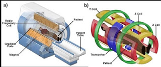
MRI MACHINE has three main parts.
(1) Scanner
(2) Computer
(3) Recording hardware
(1) Scanner
The scanner is like a big tube. This tube has a powerful magnet. The scanner has three main parts.
Static magnetic field coils-
Three types of magnetic fields can be generated in MRI MACHINE.
Fixed magnet- It has two opposite poles of permanent magnet. It consists of an alloy of iron and aluminum, nickel and cobalt. It is expensive but its operating is low.It uses a low power (0.35T) vertical magnetic field. There is no possibility of claustrophobia so it is useful for children and older patients.
Resistive electromagnet
It consists of sets of coils which run from 50 - 100kW. These coils are made of aluminum or copper. Due to resistance in it, it is very hot, so it needs cooling. This may generate a magnetic field of vertical or horizontal 0.5T. It can be switched off during emergency. It is cheap and small, the weight of this machine is about 2 tons.
Super conducting magnet
It is a large and complex machine. In this, the magnet is made of direct current coil. This coil is made of niobium-titanium alloy. It is made of a cylinder which is hollowed out from within. It has a diameter of 1m and depth of 2-3m. This coil is cooled with liquid helium that has a temperature of about 4K (-296 ℃ _. It can produce uniform horizontal magnetic fields up to 3T. It has negligible resistance, so a lot of current can be used without overheating. It is large in size and weighs up to 6 tons. It is expensive and due to the size of the tunnel, patients feel claustrophobia.
Gradient Coils-
These coils are used to generate variations in the main magnetic field. There are mainly three sets of gradient coils, one set for each direction, which is located in the X, Y and Z directions. Gradient coils produce a gradient magnetic field of 20mT / m. In this magnetic field, the location of the image slices in 3D is set in the patient by variation. The Z coil is called the Helmholtez coil, it generates magnetic fields in the crenio-codal direction in the Z direction. This field is used in the head side and more in the foot side. Hence, the proton slows down in the head side and fasts in the foot side. When RF Pulse (radiofrequency pulse) is applied to these protons, atoms whose frequencies are similar to RF resonance and generate MR signal. Hence, it is used to select the thickness of the gradient tissue. For this reason, they are also called slice selection gradients. The X and Y coils are called saddle coils. The Y-coil generates a gradient field from front to back of the patient and side-to-side through the X-coil patient. X-gradient is called frequency gradient and Y gradient is called phase encoding gradient.
Shim coils
The magnetic field generated in MRI should be uniform. Therefore, if there is an inequality, it is corrected with the help of shim coil. It imposes uniformly on the main magnetic field.
RF Coils
These coils are made in two types -
- Transmit and receive coils
- Receive only coilsRF is an electromagnetic radiation in the range of 1 MHz -10GHz. RF generates a magnetic field perpendicular to the main magnetic field. It is located near the part whose image is to be taken.
These are of the following types:
- Body coil
- Head coil
- Surface coil
- Phase array coils
- Transmit phased array coils
The body coil is a standard coil and is permanently installed in the gantry. It transmit RF and receives MR signal such as chest, abdomen. The head coil transmits RF and receives the MR signal, it is used for brain imaging. The surface coil is usually a receive only coil and is placed close to the imaging part, such as Lumber, knee and orbit. The phase array coil uses four or more receiver coils that receive different signals and then combine them together. The transmit phase array coil carries current in each element.Each has a special amplifier.
The exam is performed with the help of COMPUTER and Recording hardware and is used to store and process data. The image is shown on the display.


