Translate

BUY NOTES

BUY NOTES
Specialinvestigation लेबलों वाले संदेश दिखाए जा रहे हैं. सभी संदेश दिखाएं

HSG / hysterosalpingography in radiology

HSG / hysterosalpingography

HSG is a special type of X-ray procedure of fallopian tubes and uterus. This procedure is about 45 minutes.

Indication

  • Infertility - Infertility is the main reason for this procedure. 
  • Recurrent miscarriages - recurrent miscarriages 
  • Following tubal surgery - To check the tube opening after fallopian tube surgery. 
  • Assessment of the integrity of a caesarean uterine scar - to check for cesarean scar. 
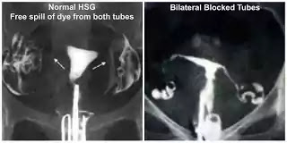
  • HSG  hysterosalpingography


  • Contraindications

    • Pregnancy- Ten Day's Rule is followed. 
    • If during the inspection of the vulva or cervix of the patient, there is no purulent discharge or if the patient has PID for the last 6 months, then this procedure is not done. 
    • This procedure is not done even if the patient is sensitive to iodinated contrast media.
    • Equipmen

      • Fluoroscopy unit with spot film device .
      • Vaginal speculum
      HSG / hysterosalpingography
      • Vaginal speculum

        • Vulsellum forceps
        HSG / hysterosalpingography
        • Vulsellum forceps


        • Uterine cannula, Leech-Wilkinson cannula, Olive of 8-F pediatric foley catheter.
        • HSG / hysterosalpingography
          foley catheter


        Patient preparation

        •  Patient should stay away from sexual intercourse during appointment and examination. Or it should use reliable contraceptive method. 
        • If a woman's menstrual cycle is regular, that is, for 28 days, then she can be given an appointment from 4th to 10th day for the procedure. 

          Preliminary Film

          • A collimated view of the pelvic cavity can be taken. 

          Technique

          •  Let the patient put supine on the examination table. The knees of the patient are flexed and the legs are abducted , and the heels are kept close to each other. 
          • Using aseptic technique, the operator inserts the speculum into the vagina and the vagina and cervix are cleaned clean with chlorhexidine . 
          • The cannula is inserted inside the cervix's anterior lip with vulsellum forceps . 
          • The contrast media is inserted into the catheter with the help of a syringe. It is taken care that there is no air bubble in the syringe and the cannula. 

            Films

            As the contrast media starts filling in the fallopian tube, the undercouch takes the image from the x-ray tube. At least 4 spot films are taken.
            • Early filling phase
            • Uterus fully distended
            • Tubal filling phase-
            • Peritoneal spillage takes the next film as soon as it appears.

            Aftercare

            It is ensured that the patient is in a very comfortable state before discharge and there is not much bleeding.
            The patient is advised that he may have bleeding from the vagina for the next 2 -3 days, which can last up to 2 weeks.
        IVP procedure in radiology

        intravenous pylography procedure

         IVP which is also done by intravenous pylography. In this series of X-ray radiographs of Kidney, ureters and Bladder system are taken.

        Indication

        This is done to detect the suspected pathology of the urinary tract, it is also used to detect the functioning of the kidney. Kidney stone and hydronephrosis are detected by this.
        intravenous pylography


        Contraindications

        In this, with the help of a catheter, the contrast media is injected into the patient's vein, sometimes the contrast media becomes allergic to the patient. So if the patient is allergic to water soluble contrast media, this test is not done. 
        Although the overall rate of these adsorption reactions is low. 
        IVP test of pregnant women is not done, as it uses non-ionic radiation, X Rage, which is harmful to unborn baby. IVP test of a patient who is known kidney disease or kidney spread is not done. Because contrast media can cause kidney function to deteriorate. 
        Elderly patients and patients who are suffering from diabetes, high blood pressure, heart disease, or are in a state of patient dehydration, are prone to kidney failure due to contrast media. 
        To prevent contrast media compaction, the patient is asked to undergo some blood tests of urea, creatine, to check kidney function.
        And their values ​​are compared with the normal range. 
        Patients suffering from diabetes who are taking metformin are asked to stop taking metformin 2 days before the examination and 2 days after the examination.

        CONTRAST MEDIA

        HOCM or LOCM 370 contrast media are used in this. 
        But the following high risk groups are ensured that they receive only LOCM contrast media - Infant Small Children and Elderly Patients. 
        Patients who are renal or cardiac failure. 
        Poorly Hydrated Patients 
        Patients with diabetes, myelometosis, sickle cell anemia. 
        Patients who have previously had severe contrast medium reaction or a strong allergic history from LOCM. Adult is given intravenously at 50ml and 1ml of contrast media for children.

        Patient Preparation

        This procedure can be done on an emergency procedure or on an outpatient basis. 
        But in most of the cases it is done on an emergency basis, because the patient brings a complex of abdomen and back pain. 
        In these circumstances this test is done without any bowl preparation. 
        In the outpatient procedure, the patient is forbidden to eat anything 5 hours before the examination. 
        The patient is kept ambulant 2 hours before the examination, so that the bowl gas can be reduced. 
        If the patient is already allergic to severe contrast media, orally methyl prednisolone 32 mg is given 2 hours before the 12 hours after the examination. 
        And then contrast media is injected.

        Preliminary Film

        For this, anti-poster image of supine full length abdomen is taken. 
        The cassette or image receptor is placed at the lower level of the symphysis pubis. 
        And the X-ray beam is placed at the midline at the iliac crist level. 
        If the radiograph is found to be satisfactory, then the procedure is continued further. 

        Technique

        The patient is placed supine on the X-ray table. With the help of 19 G Needle, the contrast media is inserted in the form of Rapidly Walls in Anti Cubital Van. 
        During this period, the stasis of the contrast media in the van may cause a pen in the upper arm or solder, which goes away from the abduction of the arm.

        Films

        In this, several radiographs are taken at different time intervals which are as follows-

        Immediate Film

        It is an anti-poster film of the renal area. The film is exposed after 10 to 14 seconds on reaching the kidney from the arm of the contrast media. 
        The film is taken to view the nephrogram. 
        During this time the contrast media is in the renal tubules, which makes the renal parenchyma opacified. 

        5 minute film

        This is also an anti-poster film of the renal area. 
        The film is taken 5 minutes after the contrast media administration, in this film it is seen whether the excretion is symmetrical or not. 
        After this a compression band is tied around the abdomen of the patient, and the balloon of the band is placed in the middle of the anterior superior iliac spine. 
        This compression band compresses the ureters in the pelvic brim. 
        If a recent abdominal surgery is performed,
        Or have a romantic romance,
        If there is a large abdominal mass, the compression band is not tied. 
        Compression bands do not apply even if the calyces appear distanced in a 5 minute film.

        15 minute film

        It is also an antero-posterior film of the renal area, in which the distended pelvic calyceal system is seen. The compression band is removed when satisfactory film is obtained. 

        Release film

        This film is taken after the release of Compression Band. 
        This suppine is the anti-poster image of full length abdomen. 
        It is also called pre void film. 
        Upon receiving this film satisfactory, the patient is asked to evacuate the bladder. 

        After micturition film

        It is also called post void film. 
        Depending on the clinical and abdominal finding of the earlier film, the film may be a full-length abdominal or bladder coned view film. In this, the tube is given a 15 degree codal angle,
        And the centering symphysis is placed 5 cm above the pubis. 
        The film describes Blader's emptying capacity. 
        It is taken to diagnose blood tumor and ureteroveseal junction. 
        The 35 degree posterior oblique film of kidney ureter bladder is taken as an additional film. 
        If only one kidney appears in the image, then 4 hour, 12 hour, and 24 hour films are also taken. After the procedure, the radiologist reviews the film.

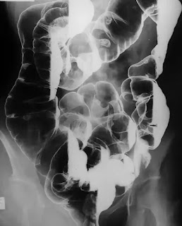
        barium enema in radiology

        barium enema in radiology

         Method

        • The ten day rule is followed in female. 
        • Single contrast - Uses 
                  Children- Because there is no need to see the mucosal pattern, so do single contrast barium enema in children.
                  Single contrast method is used to reduce intussusception.

        • Double contrast - Mucosal pattern is observed. 
        • barium enema in radiology


        Indication

        1. Change in bowel habit
        2. Pain
        3. Mass
        4. Melaena / anaemia
        5. Obstruction

            Contraindication

            Absolute

            • Toxic megacolon
            • Pseudomembranous colitis
            • Rectal biopsy via:
                       a. Barium enema is not done if the endoscopy is done 5 days before the rigid endoscope. 
                       b. Barium enema is not done if endoscopy is done 24 hours before a flexible endoscope.  

            Relative

            • Incomplete bowel preparation. 
            • Recent barium meal.

            Contrast medium

            1. Polibar 115% w / v 500 ml or more. 
            2. Air 

              Patient preparation

              There are lots of regimes of bowel preparation present. Suggested regimes-

              • 3 days before examination-
                        The patient should be given a low residue diet.

              • The day before the exam -
                       The patient should be given only fluids.
                       Picolax tablet should be given at 08: 00h and 18: 00h.


              • Exam day

                       Antibiotic prophylaxis should be given to those patients who have a prosthetic heart valve or who have a history of endocarditis.

              Preliminary film

              Preliminary plain abdominal film is not required unless-
              • Patient has severe constipation, which makes bowel preparation doubtful. 
              • Toxic megacolon suspected to be 

              Technique

              1. After heating the patient on one side of it, gently place the catheter in his rectum and connect it to the barium bottle and hand for air injection. 
              2. After this barium starts pouring in as well as intermittent screening of barium continues and as barium reaches hepatic flexure and stops barium infusion. 
              3. Now slowly pump the air into the bowel so that the barium column moves towards the caecum and produces double contrast effect. Some centers use Co2 gas for a double contrast effect, which reduces the post double contrast enema pan. 
              4. Roll the patient on its left side and bring it to the RAO position so that the bowel mocosa will coat well. 

              Films

              1. Spot film for rectum and sigmoid colon -
              • RAO
              • Prone
              • LPO
              • Left lateral of the rectum.
                    2. Spot film (Erect) for Hepatic flexure, splenic flexure and rectum -

              • LAO to open out the splenic flexure
              • RAO to open out the hepatic flexure
              • Right lateral of the rectum
                    3. Spot film for Caecum (lying) -

              • Let the patient sleep on the supine right side in the head down tilt position and take the film. At this stage, the double contrast effect occurs in the caecum. 
                    4. Overcouch film- usually exposed to the sealing tube. Large bowel is seen. (lying) -

              • Supine 
              • Prone
              • Left lateral decubitus
              • Right lateral decubitus
              • Prone with tube angled 45 ° cauded and centered 5 cm above the posterior superior iliac spine.

              Aftercare

              1. The patient should be told that due to barium, his bowel motions will be white for a few days and there may be problems in flushing. 
              2. The patient should be advised to drink sufficient amount of water to prevent barium from freezing. Laxatives can be taken if required. 

              Complication

              1. Perforation of bowel
              2. Transient bacteraemia.
              3. Changes in partial large bowel obstruction of patient due to the deposition of barium into complete large bowel obstruction.