plantar fasciitis x ray
1. Foot dorsi-plantar x-ray
Tarsal तथा tarso-metatarsal joint को देखने के लिए foot को flat position में कैसेट के ऊपर रखते हैं । तथा x-ray tube को 15 degree cranially angle देते हैं। अथवा पैरो को non-opaque pad की सहायता से 15 डिग्री कैसेट से उठाकर रखते हैं तथा x-ray beam को लम्बवत रखते हैं। यह angle longitudinal arch के झुकाव तथा tarsal bones के over showing को कम करता हैं।Position of patient and cassette
Patient को x-ray टेबल पर बैठाते है । यदि support की आवश्यकता हो तो affected side के hip तथा knee को flex करके रखने हैं। affected foot के plantar aspect को cassette पर रखते हैं। दूसरे पैर से support करने के लिये उसके knee को भी vertical position में रखते हैं । alternatively position में 15 डिग्री उठाकर रखने के लिए foam pad का उपयोग करते हैं।Direction and centring of the x-ray beam
Central ray को cuboid-navicular joint पर navicular tuberosity तथा tuberosity of fifth metatarsal के बीच मे निर्देशित करते हैं।Essential image characteristics
Tarsal तथा tarso-metatarsal joint को देखने के लिए पुरा foot examined होना चाहिये। foot की density को ध्यान में रखकर उचित kvp का चुनाव करना चाहिए जिससे कि उचित contrast प्राप्त हो सके ।Note-: tissue thickness में difference प्राप्त करने के लिए wedge filter का उपयोग किया जा सकता है।
2. Foot dorsi plantar oblique
यह projection metatarsal के alignment को tarsal के distal row के साथ मूल्यांकन किया जाता है ।Position of patient and cassette
Affected limb को dorsi plantar position से medially झुकाते है जब तक की plantar surface तथा cassette के मध्य लगभग 30-45 डिग्री का angle न बन जाएं । इस position को maintain करने के लिए foot के नीचे non-opaque angled pad को लगा देते हैं ।Direction and centring of the X-ray beam
Vertical central ray को सीधा cuboid-navicular joint पर देते हैं।Essential image characteristics
एकसमान radiographic contrast को foot density कि range में प्राप्त करने के लिए kvp select करते समय toe तथा tarsal की मोटाई के मध्य subject contrast को कम रखना चाहिए । density को एकसमान range में प्राप्त करने के लिए wedge filter का उपयोग करे । inter-tarsal तथा tarso-metatarsal joints इस projection में दिखाई देने चाहिए।
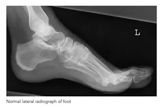
3. Foot lateral view
यह projection dorsi plantar projection के अथवा में foreign body के location को check करने के लिए किया जाता है। tarsal bones के fracture या dislocation तथा base of metatarsal fracture या dislocation को देखने में भी इस projection का प्रयोग किया जाता है।Position of patient and cassette
Foot के lateral aspect को cassette के contact में लाने के लिए पैर को dorsi plantar position से बाहर की ओर rotate करते हैं। support प्रदान करने के लिए knee के नीचे pad लगा देते हैं । foot की position को थोड़ा सा इस प्रकार adjust करते हैं जिससे कि foot की plantar surface cassette के लम्बवत हो जाए ।Direction and centring of the x-ray beam
Verical central ray को सीधा navicular cuneiform joint के ऊपर देते हैं।Essential image characteristics
यदि किसी संदिग्ध foreign body की जांच हो रही हो तो पर्याप्त kvp को select करना चाहिए । जिससे कि soft tissue की structure की तुलना में foreign body दिखाई दे सके।Note-: puncture स्थान पर metal marker को सामान्यतः foreign body की स्थिति को पता लगाने के लिए इस्तेमाल करते हैं।


