Towne's / Half axial view of skull
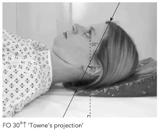
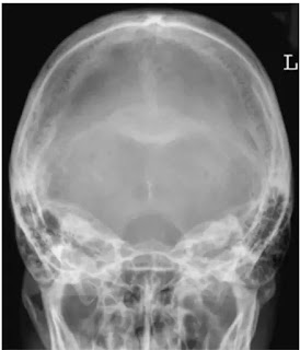
इस view को half axial या AP 30° caudal view के नाम से जाना जाता हैं। यह प्रोजेक्शन occipital bone , foreman Magnum और internal audiotory canal की किसी भी प्रकार की pathology को देखने के लिए तथा mastoid की axial position में किया जाता हैं।
Position of patient and cassette
Patient को trolley या bucky पर supine लेटाते हैं। साथ ही skull के posterior aspect को grid cassette पर रखते हैं। patient के head को इस प्रकार एडजस्ट करते है जिससे कि MSP cassette के right angle पर तथा cassette की midline के अनुरूप हो जाए। orbito-meatal base line film के लम्बवत होनी चाहिए। cassette का upper border skull के vertex से 2 इंच ऊपर रखते ह तथा शेष cassette नीचे की दिशा में रखते है।
Direction and centring of the x-ray beam
Collimated vertical x-ray beam को orbito-meatal plane से 30° caudally angle देते हैं। तथा इसके सेन्टर को midline में इस प्रकार देते है जिससे कि दोनों external auditory meatuses के बीच से गुजरे। यह बिंदु glabella से लगभग 5cm ऊपर होता हैं।
Essential image characteristics
Cassette के upper बॉर्डर को इस प्रकार व्यवस्थित करते हैं कि beam angulation के कारण skull का vertex इमेज में शामिल हो। इस प्रोजेक्शन में sphenoid bone की sella turcica foramen Magnum में project होती है। इमेज में occipital bone तथा parietal bone के posterior parts शामिल होने चाहिए तथा lambdoidal suture साफ दिखाई देने चाहिए। skull में कोई rotation नही होना चाहिए।


